Skin structure and function.

The skin is the largest organ in the body and completely covers it. It also protects against heat, light, injury and infection.
General structure
The general histological structure is composed of:
Meissner's Corpuscle (Georg Meissner): present in the touch of skin without hair, palms, plants, fingertips, lips, tip of the tongue, nipples, glans and clitoris (fine touch).
Krause corpuscles: they provide the sensation of cold.
Pacini Corpuscles: that give the sensation of pressure.
Ruffini Corpuscles: which record heat.
Merkel's corpuscles: that register to the superficial tact.
There are two types of skin:
Thin or soft skin: The thin or soft skin is that which is found mainly in the eyelids and genital areas. On the other hand, it lacks a lucid stratum
Thick skin: The thick skin is located in the lip skin, plantar and palmar, this is also characterized by having a highly developed stratum corneum compared to the rest of the skin. It is formed by corneal stratum, lucid stratum, granular stratum, thorny stratum and basal stratum.
The skin within the embryological study:
Epidermis: it has an ectodermal origin.
Dermis: has a mesodermic origin.
Biology studies the skin and divides it into 3 portions:
Epidermis.
Dermis.
Hypodermis.
While medical currents such as histoanatomy and dermatology are studied in 3 strata:
Epidermis.
Dermis.
Subcutaneous tissue.
Each layer has different functions and components that are interrelated. It is composed of: epidermis, dermis, subcutaneous tissue, and deep fascia.
skin layers
The skin is made up of two different layers. Sweat glands are distributed throughout the body. They are numerous in the palms of the hands and soles of the feet, but quite scarce in the skin of the back. Each gland consists of a series of rolled tubules located in the subcutaneous tissue, and a duct that extends through the dermis and forms a coiled spiral in the epidermis. The sebaceous glands are sac-like and secrete the sebum that lubricates and softens the skin. They open in the hair follicles at a very short distance below the epidermis.
EPIDERMIS
The outer layer is called the epidermis or cuticle. It has several thick cells and an outer layer of dead cells that are constantly removed from the surface of the skin and replaced by other cells formed in a basal cell layer, which is called the germinative stratum (stratum germinativum) and contains constantly dividing cubic cells. The cells generated in it are flattened out as they rise to the surface, where they are eliminated; it also contains melanocytes or pigment cells that contain melanin in different amounts.
t is the outer layer of the skin. Consists of two layers: The corneal layer and the Malpighi layer
The corneal layer: It is formed by dead cells that originate in the Malpighi layers. The body naturally and constantly removes many external cells from the epidermis and constantly elaborates new ones to replace the removed ones. (We are said to remove about 30,000 or 40,000 cells from the epidermis daily.) Dead cells accumulate on the surface of the skin forming a layer of keratin that must be removed to maintain good health.
In the Malpighi layer, there are cells called melanocytes that produce a pigment called melanin. The amount of melanin, which depends on race and sun exposure, is what gives the skin, hair and iris coloring of the eye.
Melanin protects the skin from the sun's ultraviolet rays and is responsible for our skin tanning in contact with the sun. The deficiency of this pigment produces albinism. Melanin is also responsible for the accumulation of blemishes, freckles, pregnancy spots, age spots and even, with excessive growth, melanone or skin cancer.
The thickness of the epidermis is generally very thin, although there are areas with different thickness. Thus, while in certain areas such as the soles of the feet or palms of the hands, it can measure 1.5 mm, in other places, such as the eye contour, is less than 0.04 mm. The epidermis is the external barrier that protects us from external aggressions and maintains the appropriate level of internal fluids, also allowing, through its permeability, that some of them can go outside.
.gif)
DERMIS
The inner layer is the dermis. It is made up of a network of collagen and elastic fibers, blood capillaries, nerves, fatty lobes and the base of hair follicles and sweat glands. The interface between dermis and epidermis is very irregular and consists of a succession of papillae, or finger-like projections, which are smaller in areas where the skin is thin, and longer in the skin of the palms of the hands and soles of the feet. In these areas, the papillae are associated with elevations of the epidermis that produce undulations used for fingerprint identification. Each papilla contains either a capillary loop of blood vessels or a specialized nerve termination. Vascular bonds provide nutrients to the epidermis and outnumber the neural papillae, in an approximate proportion of four to one.
It is the layer covered by the epidermis. It consists of two continuous layers. In it we can find:
The sweat glands, in the form of a spiral with a tube that is projected to the outside, constantly produce sweat that exits the dermis through the pores. With sweat we eliminate toxins and regulate body temperature.
Sebaceous glands, in the form of a sac, produce sebum or fat towards the dermis. The function of sebum is to lubricate and protect the skin. The sebum and sweat combine to form a layer that protects the skin and makes it impermeable to water.
Adipose cells: are found in the lower part of the dermis. Its function is to pad the body protecting it from shock and providing heat.
Hair follicles, which, in the form of a tube, are born from fat cells and continue to the epidermis. The hair is produced inside. Each hair follicle is lubricated by a sebaceous gland which is the one that provides fat to the corresponding hair. This grease polishes it and protects it from moisture.
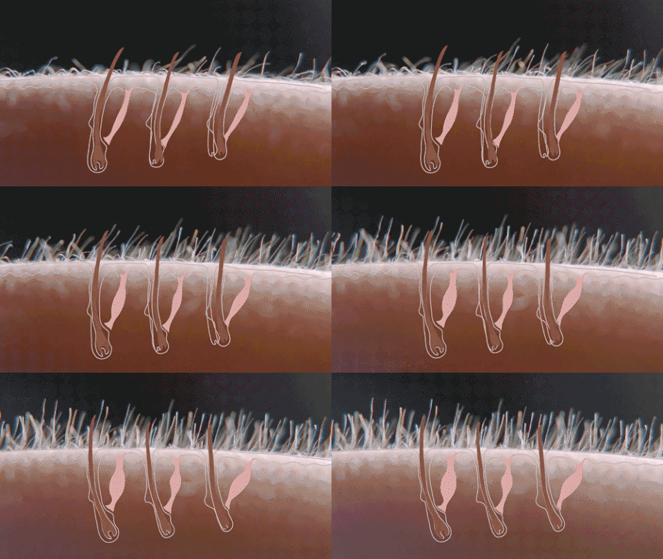
The hairs are held by elevating muscles that when contracting erizan the hair. This is what happens when we feel certain tactile sensations, or in the face of fear, cold, and so on.
Blood vessels that supply the different cells of the skin through the capillaries
Collagen and elastin fibers: They are found in the deepest layer of the dermis. Its function is to keep the skin smooth, elastic and youthful.
The nerve fibers responsible for sensations. Sensations are formed when the receptors send the perceived information to the nervous system. These receptors receive different names according to the type of sensation they capture. Thermoreceptors are able to identify sensations of heat or cold (thermal sensations), mechanoreceptors capture the weight of objects (pressure sensations) and the shape, texture, size, etc. of objects (tactile sensations); nociceptors capture pain (painful sensations)
Nerve fibers can be free, with bare sensory fibers (14) or covered by connective tissue. They end up in bumps called corpuscles. We have the following corpuscles:
Paccini corpuscles: They appear encapsulated. They are made up of a series of spiral layers formed by flattened connective tissue that resemble ce bollas in shape... They are responsible for collecting vibrations and pressure, so they are very abundant in the hands and feet.
Ruffini's Corpuscle: They have an elongated shape and appear in the deepest part of the dermis. Its function is to detect skin and subcutaneous tissue deformations. They also capture heat. They are more abundant in the hand by the top face.
Meisner's corpuscle: Egg-shaped, appear mainly at the tip of the toes and feet. They respond to gentle touches on the skin. They are able to quickly detect the shape of objects and their textures.
Krause Corpuscle: They appear encapsulated in the deepest level of the skin. They are similar in shape to Pacini's corpuscles, although they are smaller and somewhat more rounded. They are believed to be able to detect the cold. They can be found in the mouth, nose, eyes, tongue, genitals, etc
Here I show you an example when the skin is mistreated by needles where the person's desire is to be tattooed when in fact it is a damage to the skin, so its treatment of steemians healing.
Functions of the skin
The skin is the largest organ of our body. The skin of our body externally, the internal organs, muscles and bones, making the whole body appear as something compact. Its thickness depends on the area it covers, so in the eyelids is very thin and only half a millimeter thick, while in the soles of the hands and feet is about 4 mm.
It is an organ that fulfils fundamental functions in the organism. It is considered a huge gland that covers the entire body, separating and uniting the inner and outer world.
It fulfills multiple functions:
PROTECTION: Protects our body from the outside world. For example, trauma.
THERMOREGULATION: Regulates the constant temperature of 37 degrees that the individual needs. This is why it is called the peripheral heart.
SENSITIVITY: For this function is that we feel heat, cold, etc.... This is why it is called the peripheral brain.
DEPOSIT: It is a reservoir of multiple substances such as: minerals, fatty substances, organic substances, hormones, vitamins, etc....
EMUNTORY: It is the elimination of different substances through sweat and sebaceous secretion.
ANTIMICROBIAL: It is the first great defense of the organism and acts as a natural barrier. If this barrier is broken, infection occurs.
MELANOGENIC OR PIGMENTATION: Melanogenic cells are found in the basal layer of the epidermis, which produce melanin, which gives the skin different tones. That's how we have the different races:
White Race: Less melanin and less protection.
Yellow Race:
Black Race: More melanin and more protection.
These pigments protect us from the sun's rays. Albinos do not have pigments, so they must avoid the sun, which will cause major burns and can lead to skin cancer. Pigmentation intensifies in summer and decreases in winter. The white and sensitive skins of blond, redheaded and children's skin were to be protected with sunscreen and sunscreen in the summer.
BIOLOGICAL DETERIORATION

Wrinkles
Wrinkles are caused by physical-chemical alterations that lead to skin aging. As time passes, three important elements for the skin are gradually lost:
Collagen (the protein fiber that gives firmness to the skin), which causes it to become thinner and weaker.
Elastin, responsible for elasticity;
Glycosaminoglycans, moisture retention.
Moreover, the sun, tobacco smoke and pollution can also speed up the process.
Source of information
- https://www.myvmc.com/anatomy/human-skin/
- http://www.bbc.co.uk/science/humanbody/body/factfiles/skin/skin.shtml
- https://www.ncbi.nlm.nih.gov/pubmed/11250091
- https://www.proteinatlas.org/humanproteome/skin


.gif)
