Steemit Learning Challenge-S21W2: Physical Therapy Intervention: Knee Osteoarthritis
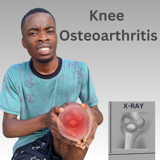
edited using canva
Let's assume you're a motorcycle owner and the bike is new with new tires. The tire is there to help or support movement of the whole body of the bike and provide support to it. With these new tires on the bike, it has what we call threads or hairs seen on the body of the bike, or maybe it has some structured design that prevents it from colliding with the floor, creating room for friction.
It's normal that with time, those hairs on the body of the tire will grow thin or smaller, and the tire due to too much driver will conceal those structured designs and make the tire look smooth. When the tire is smooth like this, there's no friction with the ground for support.
The tire becomes smooth and can slip on slippery or bad roads. You begin to feel every rough patch on the road, which makes driving very uncomfortable. The knees are just like that tire that protects the bones from rubbing against each other.
When there's the presence of what we call osteoarthritis, which causes wear and tear of the joint cartilage, the cartilage behind the knees wears down just as the tire wears and becomes smooth. When this wear and tear happens, the knee starts to join together between bones, making it stiff, swollen, and painful. With this, you should have a glimpse of what I'm to talk about since Knee osteoarthritis is a medical disorder term.
What's knee OA? Write in your own words after getting knowledge from the lesson post. |
|---|
The knee is a part of the body that is found before the upper forelimb. It is that connector in bone form that helps the legs and body move. Without your knee, your legs are gone because there are no legs without knees. So the knee is very important in movement, which consists of cartilage that balances the joints and bones, making it flexible to move back and forward.
The knee is structured in such a way that the thighbone, shinbone, and kneecap make up the joint found in the knee. It also consists of an elastic tissue that covers the ends of the bones, which allows for smooth movement and reduces friction.
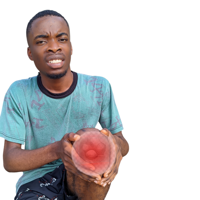
This elastic tissue is called the Cartilage. It also has ligaments and tendons that stabilise the knees and synovial fluid that lubricates the knee constantly, reducing friction that may cause clashing of bones.
OA, as abbreviated, means Osteoarthritis. It's a joint word and not a compound or separate word. It is a form of arthritis that tackles the knee, causing stiffness, reduced mobility, and pain. So if I'm joining the two words together, we have knee osteoarthritis.
Knee osteoarthritis is a joint disease that is degenerative and affects the cartilage, tissues, and bones in the knee. The joint found in the knee bears a lot of weight and all the forces of movements in running, jumping, walking, and lifting heavy objects.
That's why most times the knees are affected when you jump and not land well or when you run and slip; your knee shifts. The stress that results in too much force of movements by the knee makes it prone to having that medical disorder. Factors like ageing, injury, and heredity cause this medical disorder of the knees.
As one ages, the knee cartilage becomes stiff and can likely cause this medical disorder. Most old people today, especially in my region, face this problem which affects their movements. You'll see them find it difficult to walk well. Sometimes I think if this is a trait that all old people must have leg problems.
Injury too can cause this indirectly. Our parents will always earn us against playing round play or having wounds. Taking care of your body now tells how your old age will be. If you have a fracture in your bone now, it's possible you won't be able to move freely when you're old, as you'll either be faced with this form of arthritis or other bone disorders.
This medical condition is sometimes generally related, as it may be a trait in your family. Obesity, excess weight lifting, and situations that involve standing for longer hours without moving may make your knees susceptible to such a disorder. I've now seen the reason why my knee feels so much pain if I stand for too long in a two-hour lecture.
Symptoms
The symptoms are mostly physically seen, which includes inflammation, which causes swelling in areas close to the knee. You'll also hear your knee grind or crack when you move, run, or stand for too long. Consistent pain in that area when moving can be a major symptom. These are some observable symptoms that give us a clue that someone has knee osteoarthritis. Internally, you'll find your knee very stiff and hard. This is another symptom not observable. This medical order has stages.
TYPES
There are different types of knee osteoarthritis, even though knee osteoarthritis is a category I see as arthritis. The below explains best.
- Primary osteoarthritis: This is a type of knee osteoarthritis where the exact cause is not known. It usually comes with ageing where the knee is brittle. This type affects both knees in older adults.
- Secondary _: This is a type where there's a specific known cause for the disorder. It can happen in young people and mostly affect one knee. Conditions like diabetes and obesity usually trigger it.
- Post-Traumatic Osteoarthritis,: This is a form of secondary knee osteoarthritis that develops when the knee is seriously injured, such as a fracture. Symptoms may not be imminent and may develop months or years after the fracture or injury.
We have another type that occurs when there's inflammation that affects the knees and yet another that occurs when there's a breakdown of the cartilage and wear of the bone. These are the types of knee osteoarthritis that have stages it develops. The two major types are the Primary and secondary osteoarthritis. The stages include;
Normal
Minor
Mild
Moderate
Severe
- As we can see in the first stage, there's no osteoarthritis. The knee is very healthy, and cartilages and joints are functioning properly.
- Minor stage: is a stage where there's a little wear in the knee cartilage. It's called minor because there's no major wear. It doesn't affect the function of the joint, but the patient will experience mild pain as a result of stiffness when there's a prolonged, weighty activity done.
- Mild stage is a stage where the cartilage of the knee begins to wear, leading to pain occasionally. This breakdown is slightly done; that's why it's termed a mild stage.
- Moderate or average stage results when the cartilage has completely worn down in such a way that there's a curve in the knee. In this stage, the pain and stiffness are increased compared to those of the mild. Moving becomes difficult, and one in this stage may experience a consistent clash in his bones as he moves.
- Severe stage occurs when the cartilage is not there to act as a supportive elastic tissue and the bones are clashing against each other. You'll experience chronic and severe pain that restricts mobility. There's always inflammation in the knee region at such a stage. These are the five stages that define the knee osteoarthritis medical disorder.
So let's assume you have any of the stages. How would you know, and what can you do to recover? The following diagnosis gives a preview.
How would you diagnose a knee OA? Any clinical investigation or assessment tests? |
|---|
There are various ways to check if a patient has the medical disorder knee osteoarthritis. It can be through medical records, physical examinations, clinical and physical tests, etc. The information below explains everything in detail. The first thing to do when you have a patient is to check his medical history or records.
- Talk with the patient to gather symptoms and his medical records or history to understand the patient's experience and how his symptoms affect his daily life. You can do this by asking questions like;
Do you feel pain in your knee region often, especially when you're walking or climbing a stair? Is your body always stiff when you sit for a while and stand up?
If his answer to the questions is an affirmation, you can be certain that he has knee - **Talk with the patient to gather symptoms and his medical records, or as people suffering from it feel more pain in the knee after an activity is done. You can also ask for the duration as to how long he has experienced this.
You can ask, if necessary, if any member of his family has experienced such before. This disorder can be genetic as well. Once we are done interrogating the patient, we can move on to a physical examination of the patient to confirm these pain areas.
Physical Examination
A physical examination is very important though, as it helps you as the doctor confirm these pain areas. You can do this by observing if there's any swelling in the knee or deformities like a fracture, joint knees, or something else. After observing, you can gently rub the joint line to check warmth and tenderness, which is a "palpation technique.
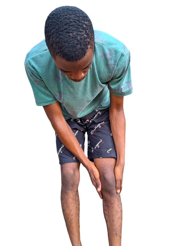
The aim of this physical examination is to determine physical changes through observations, range of mobility, stability, and testing for pain.
You, as the doctorwill place your hand over the knee of the patient and they will bend it. You're trying to check if there's grinding or cracking when bending. If this happens, the joint is rough. After placing your hands to feel the knee for grinding, you check how well he can move his knee and leg, which will determine the range of motion of this person.
You'll tell the patient to bend and straighten his knees five times. If he finds doing so very painful, it's evident that he's got knee osteoarthritis. You can ask the patient to walk for 5 seconds to see if he's limping or not. After these physical examinations to check and confirm pain areas, the doctor will allow the patient to carry out some physical assessment test that involves motion.
Assesment Tests
These are tests that are done physically by the patient with the help of the doctor to check mobility. The first assessment test to check is what we call the TUG Test.
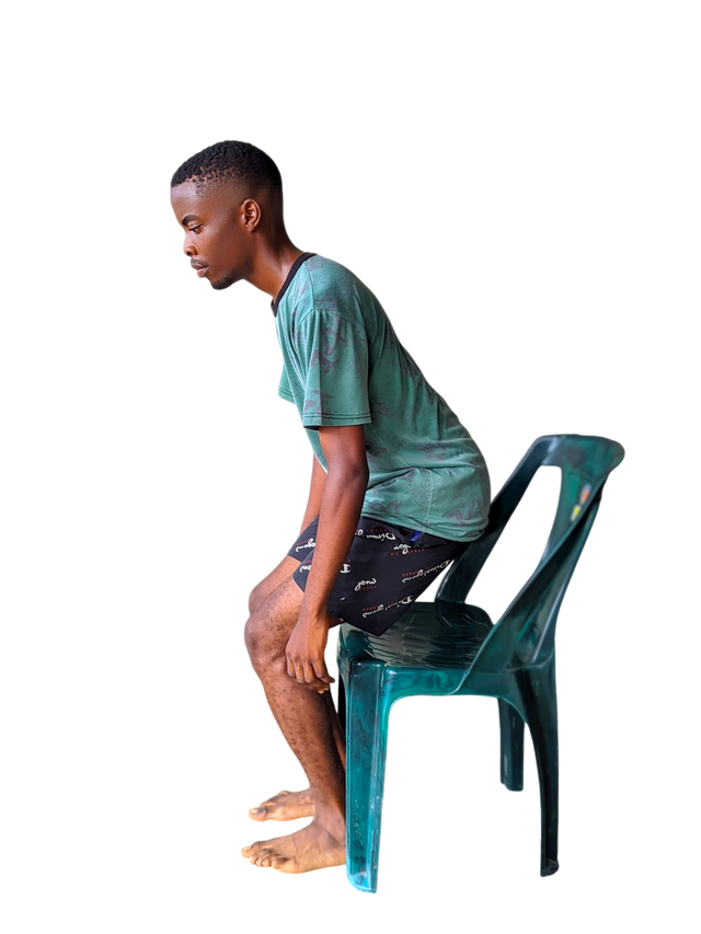
- TUG Test is carried out by telling the patient to stand up from his chair, walk 5 meters, and then return to his seat. You're to allow the patient to do this all by himself to observe if there's balance in mobility or not. He's to do this for 30 seconds to 1 minute as he walks, turns around, and then sits down back in his chair. The second one involves what we call a speed test by telling the patient to sit and stand consistently for 30 seconds or less.
- Sit-to-Stand Test: The patient is told to stand up and sit down as quickly as possible. His doing this helps the doctor assess his lower body strength and his ability to endure lifting his body while the knees are bent and strengthened.
 | 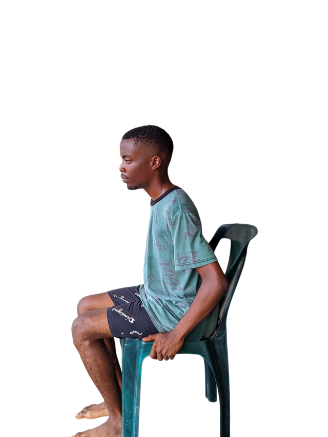 |
|---|
- The single-leg stance test can be employed to check for stability and balance, which is mostly active in the knees. The knee places its sole focus there. You can tell him to stand on one of his legs for as long as 1 minute.
- Tandem walk test is achieved when the doctor tells the patient to walk on a straight line with one of his feet replacing the other in a forward motion. The left leg's heel touches the toe of the right leg, and then the right leg's heel touches the toes of the left leg. This helps to check balance, as the knees usually come into contact when carrying out this exercise.
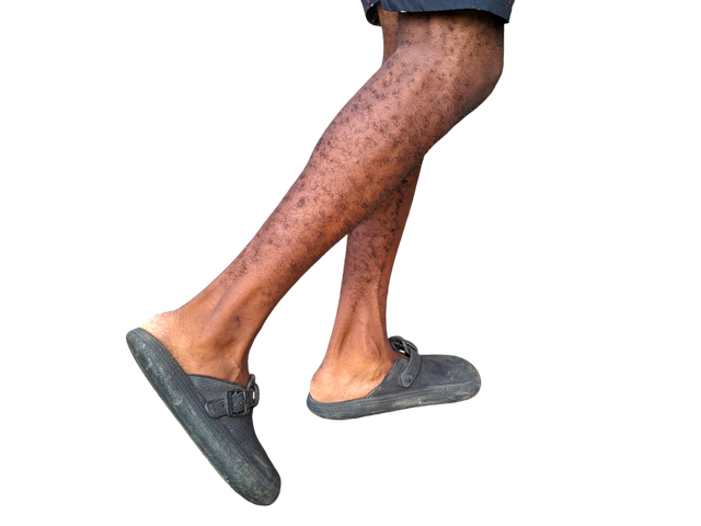
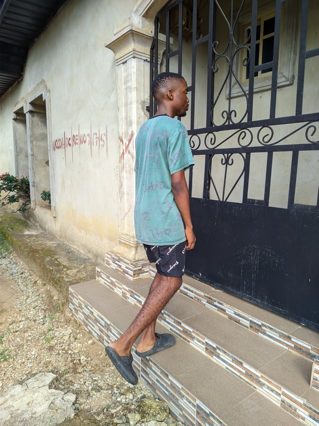
- Stairs climbing: This involves the knees and legs as the knees are bent while climbing the stairs. It helps the doctor to assess if there's difficulty lifting the legs on the stairs. These are some physical assessment tests that can be carried out.
 |  |
|---|
Laboratory Tests and Imaging
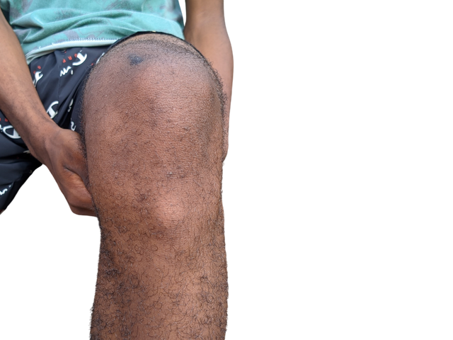
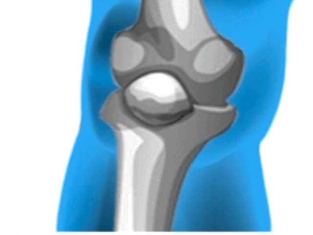
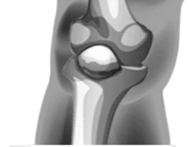
The use of imaging is to provide a clear outlook at the internal structure of the forelimb of the knee, which would display the physical effect of osteoarthritis. The use of X-ray can be essential in showing the bone and changes in the cartilage. The x-ray reveals narrowing space between the joints, spurs in the bones, and thickening of the bone below the cartilage of the knee.
 | Imaging the knee |
|---|
 |  |
|---|
pictures gotten from an x-ray of my picture using the x-ray app
It doesn't give full details compared to the use of an MRI. This provides a detailed image of soft tissues in the knees, which can be used to check if there's any ligament damage. This MRI is not necessary, though, but is helpful in providing details.
The use of ultrasound in detecting inflammation in the joints or joint effusion can be helpful when giving injection as it guides the direction of the injection to avoid complications.
A CT scan is helpful when details of the bones are needed. These are a few imaging studies that can be carried out to ensure this.
Try to practice at least 4 exercises that you have learnt from the lesson. Share images, gifs or videos while practising. |
|---|
These exercises were mindful and helped me a lot in strengthening my legs. I'll keep on trying it every day to be sincere. I love the program. I don't have a straight leg, which doesn't mean I have knee osteoarthritis because my knee doesn't hurt me often. It's only when I hit it that the pain goes away for a few days. The first exercise I did for this contest was Knee to chest
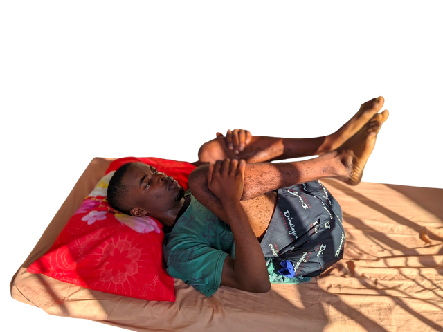
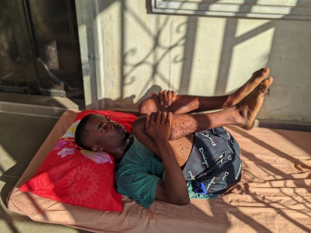
- Knee to chest: I did this by first laying on a soft bed with my pillow to support my head from being flat. I then folded my legs to meet my chest and held them with my hands while trying to release them, and then brought them to meet my chest again. The bending of my knees really helps straighten it, and I didn't feel pain doing the exercise. It was just the cracking sound of releasing and bringing my legs together with my knees bent.
 |  |
|---|
This is a practical video that explains how I did this best. This exercise helps to increase the ROM of my knee joints. I did this exercise for more than one minute to strengthen my knees the more. It was indeed helpful, I must admit.
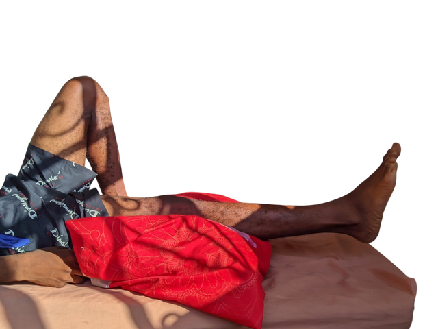
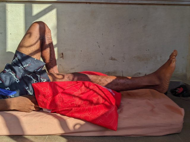
- Knee Flexion: This is an exercise for the knee's joint to improve and also increase the ROM of the joint. It's also an exercise for strengthening the leg's muscles and muscles around the knee. I did this exercise for more than one minute, and I laid on a small bed with my pillow below my leg in the absence of a towel. The pillow was made to raise my lower legs and knees.
 |  |
|---|
While the knees are placed on the pillow and my lower legs on the foam. All I did was to raise my leg up and then suspend it on the air in a horizontal pattern for 5 seconds before bringing it down. I did this continuously while giving more attention to my knee.
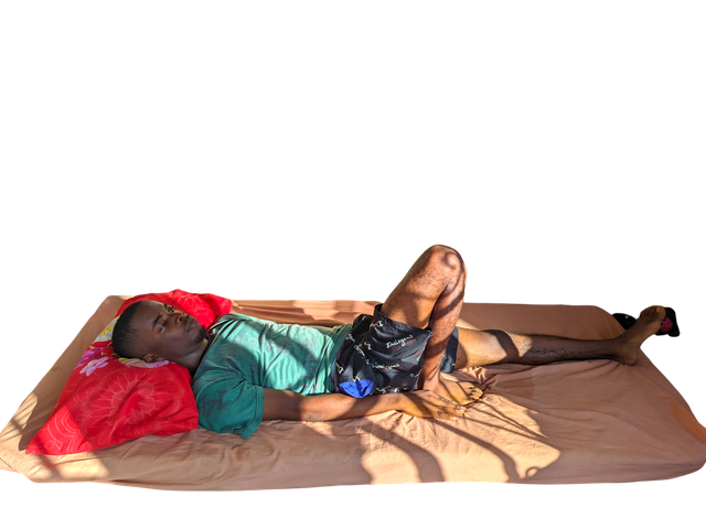
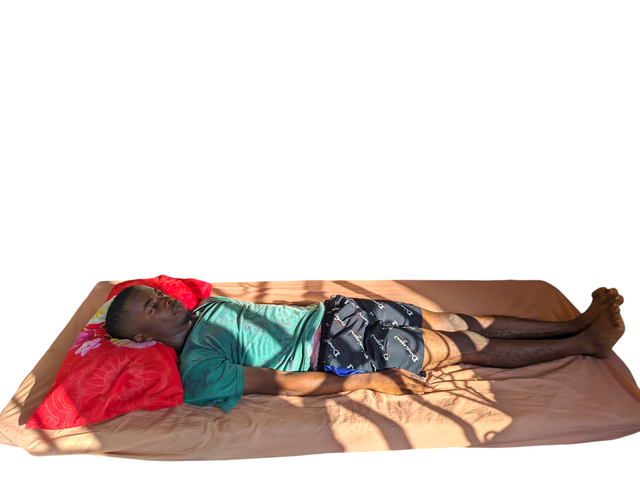
- Heel Slide Exercise: This is an exercise that helps prepare the patient to walk as the knees and legs are brought in contact, strengthening the muscles of the legs. I did this by lying on a foam, stretching my legs as if I'm lying dead, and then moving one of my legs to my bottom. I did it by dragging it. My knee and legs felt the impact, I must admit. I did this exercise for more than a minute, as shown in the video.
 |  |
|---|
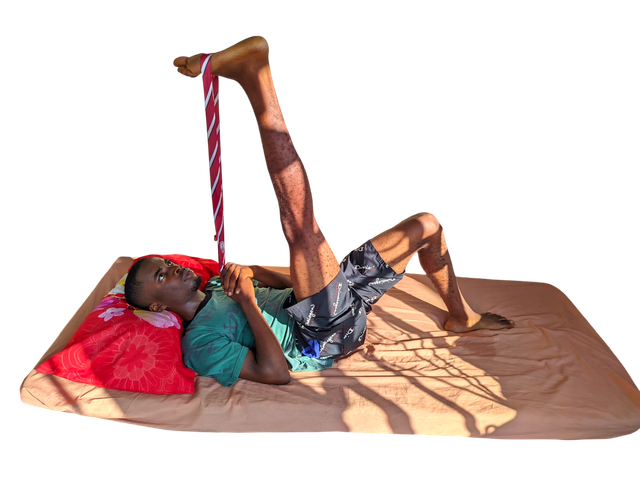
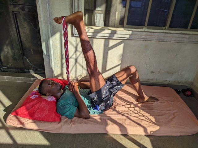
- Calf and Hamstring Exercise: This is an exercise done to improve the shortened muscles, and I did this for more than a minute. The exercise was done by laying on foam and then placing a tie on my feet while my legs are upright in a vertical position. The to-and-fro movement did help strengthen my leg muscles and knee.
 |  |
|---|
Share your review after performing these exercises either as a healthy individual or patient. |
|---|
I'm healthy and don't have this medical disorder. My doing this exercise is not because I have a disorder. I want to improve my legs so that it won't affect me later in the future when I advance in age. Exercise keeps the body fit, and these few exercises will help improve my legs muscles and my knee's joints as well.
Before the advent of this course, I've been working on this exercise, and doing it every week has helped me gain balance and the ability to move in a flexible and not rigid way.
I invite @radleking, @artist1111 and @walictd
Cc,
@ashkhan
https://x.com/bossj23Mod/status/1853926525692674330?t=ZaysJetzolt3101PtLmzlQ&s=19